For most patients, ultrasound is a relatively painless procedure. Conventional ultrasound is safe, low-cost, non-ionizing, and one of the most commonly used imaging modalities worldwide. In a typical exam, a clinician presses an ultrasound probe with gel on the skin surface to generate an image at the desired location. The probe emits sound waves into the tissue and various features such as muscle, fat, blood vessels, and bones reflect sound back to the probe, which records the reflected signals and produces an ultrasound image. Since the probe must make contact with the skin surface to transmit and detect ultrasound, the probe's orientation on and compression of the skin surface creates contact sensitive images. This contact sensitivity causes unaccounted image variations that restricts ultrasound performance. Clinicians imaging the same region at two different times will typically produce two different images, rendering quantitative tracking of tissue change over time impossible. Furthermore, skin contact may be limiting in situations where patients don't tolerate probe contact well, such as neonates, trauma/burn victims, or surgical patients.
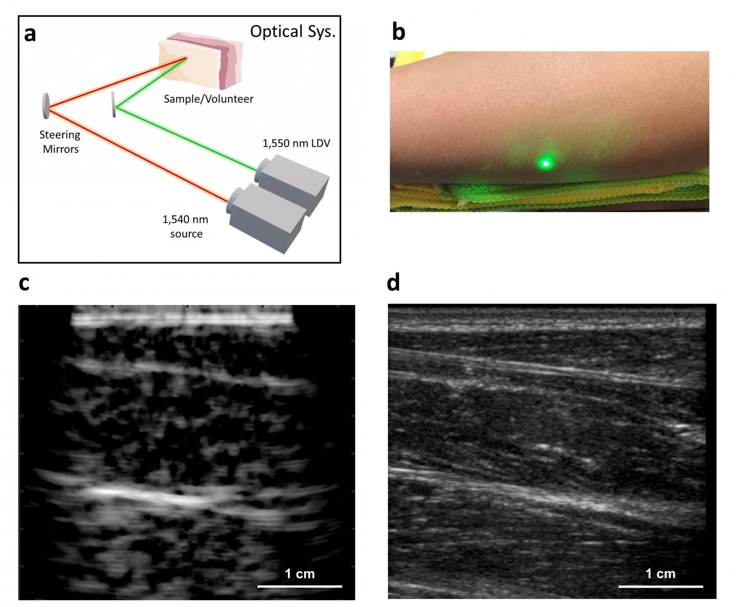
In a new paper published in Light Science & Application, Xiang (Shawn) Zhang and Brian Anthony from the Mechanical Engineering Department at Massachusetts Institute of Technology, in Cambridge, United States, developed an alternative approach to conventional ultrasound that does not require direct contact with the skin surface. The new laser ultrasound technique uses eye- and skin-safe lasers to remotely generate and detect ultrasound on the skin surface. The transmit laser sends a light pulse which is rapidly absorbed on the skin and converted into sound waves via the photoacoustic effect - the generation of sound by light. The generated sound waves interact with tissue identical to conventional ultrasound and the reflected signals are detected by a laser interferometer on the skin surface. The lasers are moved across the skin surface to produce an image. The system was successfully tested on human subjects and showed that laser ultrasound is sensitive to tissue features presently detected using conventional ultrasound. The new technique can image at centimeter depths which is significantly deeper than other optical ultrasound techniques and is comparable to imaging depths of modern clinical ultrasound,
These first laser ultrasound image results are very encouraging. "We're at the beginning of what we could do with laser ultrasound. Imagine we get to a point where we can do everything ultrasound can do, now at a distance. This gives you a whole new way of seeing internal structures, without making contact with the patient." The researchers hope to improve upon the current technology and push laser ultrasound toward future clinical use. "As related technological fields advance, one can imagine designing a laser scanning system that can remotely and volumetrically image a patient without ever disturbing or making contact."
EurekAlert!, the online, global news service operated by AAAS, the science society: https://www.eurekalert.org/pub_releases/2019-12/cioo-fnl122419.php