Direct atom-resolved imaging of magnetic materials
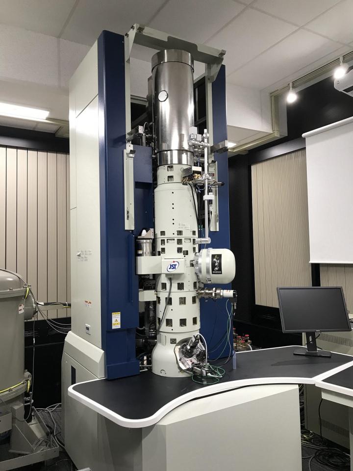
Under the JST-SENTAN program (Development of System and Technology for Advanced Measurement and Analysis, Japan Science and Technology Agency), the joint development team of Prof. Naoya Shibata at the University of Tokyo and JEOL Ltd., has developed a revolutionary electron microscope that incorporates newly designed magnetic objective lenses, and achieved direct, atom-resolved imaging of materials with sub-Å spatial resolution, with a residual magnetic field less than 0.2 mT at the sample position. To the best of our knowledge, this is the first time that such a goal has been achieved.
In the 88 years since the seminal invention of the transmission electron microscope (TEM) in 1931, researchers have continually pursued better spatial resolution. The design of magnetic objective lenses with smaller lens-aberration coefficients has been necessary, and aberration-correcting lens systems for scanning TEM (STEM) have achieved sub-Å spatial resolution.
One critical disadvantage of current magnetic condenser-objective-lens systems for atomic-resolution TEMs/STEMs is that the samples must be inserted into very high magnetic fields of up to 2-3 T. Such high fields can severely hamper atomic-resolution imaging of many important soft/hard magnetic materials, such as silicon steel, because the strong field can greatly alter--or even destroy--the material's magnetic and sometimes physical structure. Recently, the development of new magnetic materials has advanced rapidly. As atomic-scale structural analysis is key to the abovementioned technology, a solution to this problem has long been required.
The joint team has developed a new magnetic-field-free objective-lens system, containing two round lenses positioned in an exact mirror-symmetric configuration with respect to the sample plane. This new lens system provides extremely small residual magnetic fields at the sample position while placing the strongly excited front/back objective lenses close enough to the sample to obtain the short focus length condition indispensable for atomic-resolution imaging. Consequently, the residual magnetic fields generated near the sample center are much <0.2 mT, which is 10,000 times smaller than the value in conventional magnetic objective lenses used for atomic-resolution TEM/STEM imaging.
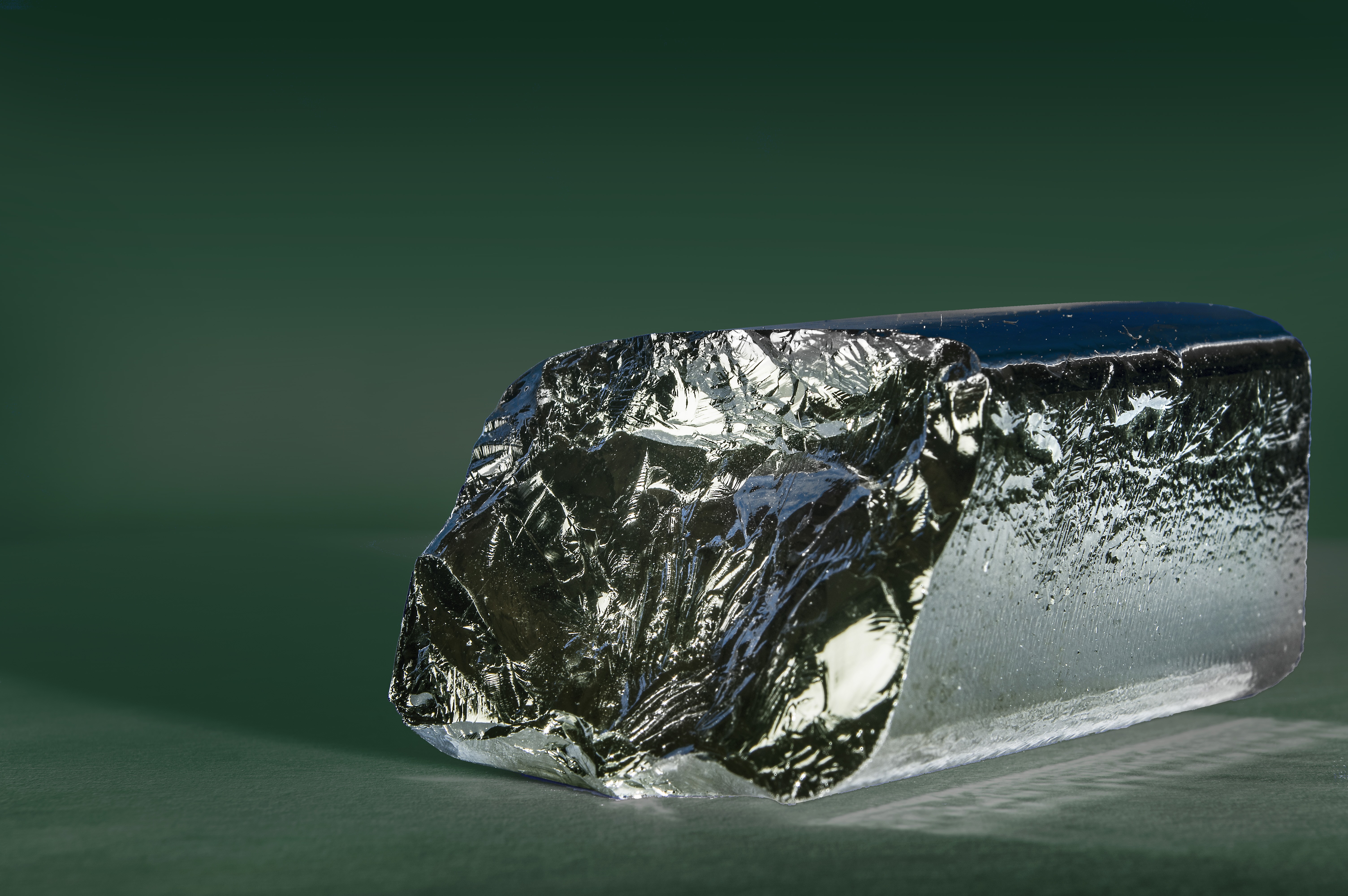
The joint team has used this new system to observe the atomic structure of a grain-oriented silicon-steel sheet, which is one of the most important soft magnetic engineering materials. This sheet is used as a core material for electric transformers and motors, and its atomic-resolution characterization of individual defects has long been sought. Using the newly developed lens system, the resolved atomic structure of the silicon steel was clearly observed, and direct, atom-resolved imaging in a magnetic-field-free environment was realized for electron microscopy, enabling unprecedented atomic-level structural characterization of magnetic materials.
The newly developed electron microscope can be operated in the same manner as that of conventional TEMs/STEMs. It is expected to promote substantial further research and development in various nanotechnology fields.
EurekAlert!, the online, global news service operated by AAAS, the science society: https://www.eurekalert.org/pub_releases/2019-06/jsat-aie061119.php